
A team of radiologists at the Ghent university hospital has achieved a world first by creating three-dimensional CT bone scan images without using dangerous radiation.
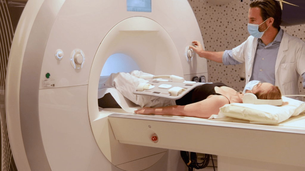
There are two main types of scanner used in medicine: CT and MRI.
MRI scanners use harmless radio waves, but are more useful for scans involving soft tissue rather than bones.
The CT (or CAT) scan is better for scanning solid structures like bones, but it uses X-rays, which have dangers of their own.
Working together with the Dutch software company MRIguidance, the team spent three years working to achieve a sort of hybrid of the two – the patient is subjected only to the benign MRI waves, while the software is able to convert those scans into CT-quality images.
The breakthrough earned the researchers the cover of the journal Radiology, where the research was published.
“Thanks to the Dutch software, we can use artificial intelligence to convert MRI images of bones into accurate CT images in 3D in an hour and a half,” explained Lennart Jans, professor of radiology at UZ Gent, who has claimed an international first.
For the patient, all that is required is for them to stay in the MRI scanner for an extra four minutes.
“The big advantage of this new technology is that you no longer need a traditional CT scan and that harmful radiation is not required,” Prof. Jans told De Tijd. Another advantage is that the modified scan allows top quality images of both bones and soft tissue, including muscles and tendons.
Every year, two million CT scans are carried out on patients in Belgium, each one carrying the risk of causing cancer. For that reason, doctors tend not to prescribe a CT scan unless absolutely necessary, even in cases of bone injuries, where they have real utility.
“The result is that we miss important information needed for a good diagnosis and correct treatment,” he said. “This new technique transcends the drawbacks of the existing scans and is a game changer.”
The technique has been tested in recent months at UZ Gent in patients with inflammatory rheumatism of the sacral joints, where the hips join the spine, a condition known as ankylosing spondylitis.
“Surgeons can now save time in surgery planning, and patients only have to come to the hospital once,” Prof. Jans said. “This is also a saving for health insurance. Within a few years this technology will become the standard.”
Alan Hope
The Brussels Times